Skip to Content
Schedule A Call

QSS DICOM/PACS Viewer

DICOM Viewer Software for Faster, Clearer Reads, Anywhere

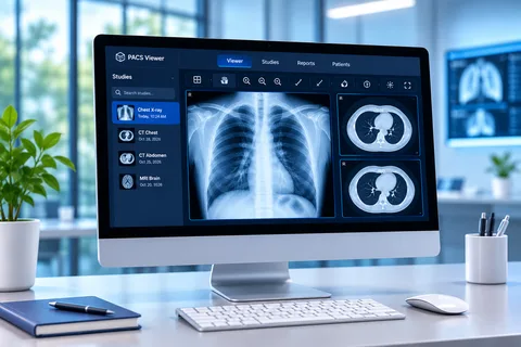
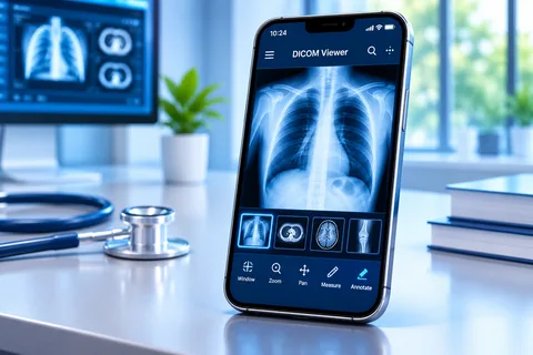
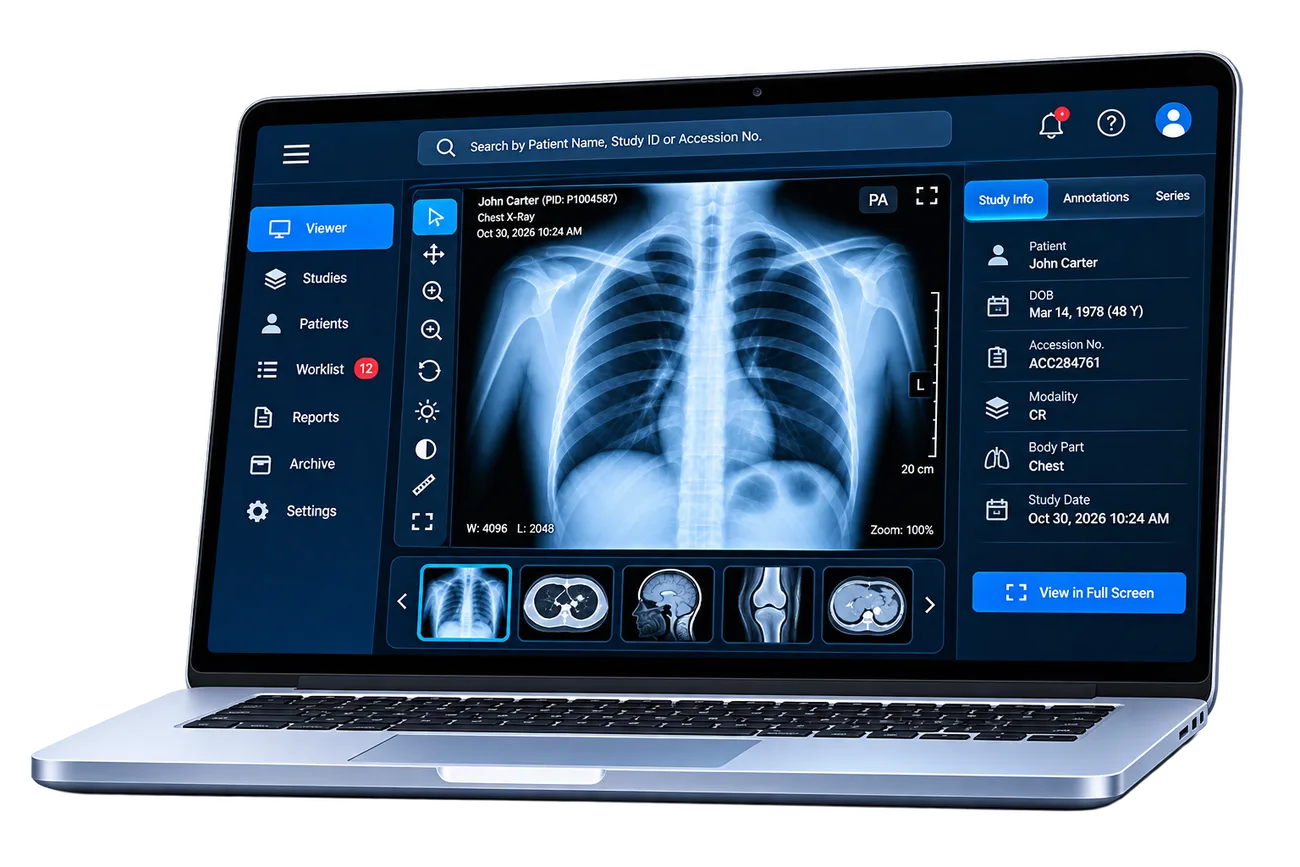
The QSS DICOM/PACS Viewer is DICOM viewer software that opens CT, MRI, X-ray, ultrasound and PET studies in a browser, on the desktop or on a mobile device. Radiologists and clinicians get diagnostic-quality images, measurement and annotation tools, MPR and 3D, and structured reporting in one place, connected to your PACS, RIS, HIS and EHR.

Multi-modalityCT, MRI, X-ray, US, PET
MPR and 3DVolume rendering built in
PACS, RIS, HIS, EHRHL7, FHIR, DICOM
HIPAA-readyBuilt by a CMMI Level 3, ISO 27001 team

TRUSTED PARTNERS

Medical imaging software from a team that has shipped software since 2010

The QSS DICOM/PACS Viewer is built and supported by QSS Technosoft, a US-headquartered medical software development company in Bloomington, Minnesota, with delivery centers in the USA, India and Dubai. The same engineers who build the viewer also integrate it with the systems your clinicians already use.

POWERING BETTER CARE TOGETHER

400+Projects delivered to production
200+Clients served
20+EHR/EMR integrations delivered
4.9/5Client rating on Clutch

What it solves

Where imaging workflows slow down, and what changes with the QSS viewer

Most delays in imaging are not in the scanner. They are in getting the right study, with the right priors, in front of the right person, and then getting the report back into the chart. Here is where radiology and clinical teams lose time, and what the viewer does about it.

Problem

Images are locked to one workstation in the reading room

How we solve it

A web DICOM viewer that opens studies in a standard browser, plus a desktop app and mobile access, so authorized users can read from the hospital, the clinic or home.

Studies reach the reader, not the other way around.

Problem

Referring physicians wait for CDs or phone calls to see images

How we solve it

Secure, role-based links and EHR launch let referring doctors open the images and the report from the patient record.

Fewer CDs, fewer calls, faster decisions at the bedside.

Problem

Every modality needs its own viewer

How we solve it

One viewer for CT, MRI, X-ray, ultrasound and PET, with the same tools and layouts across modalities.

One tool to learn, support and secure.

Problem

Complex cases need 3D, but 3D lives on a separate workstation

How we solve it

MPR and 3D volume rendering run inside the same viewer, on the same study, without exporting data to another system.

Advanced views when you need them, in the workflow you already use.

Problem

Reports are typed in one system and images sit in another

How we solve it

Structured reporting templates sit beside the images, and the finished report flows back to RIS and EHR over HL7 or FHIR.

A cleaner report and fewer re-keying errors.

Problem

Sharing images safely is hard to prove to auditors

How we solve it

Role-based access, encryption in transit and at rest, and a log of who opened which study and when.

Image access you can explain in a HIPAA review.

Want to see these workflows on your own studies? We will run a live demo with anonymized sample data from your modalities.

Talk to Our US Team

Features

Everything a modern DICOM viewer should do, grouped the way you work

Explore the healthcare software areas where we help organizations build, connect, modernize and scale.

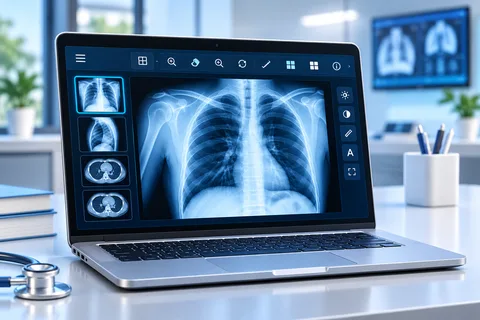
Core Viewing

Fast, diagnostic-quality viewing for every common modality, in the browser or on the desktop.

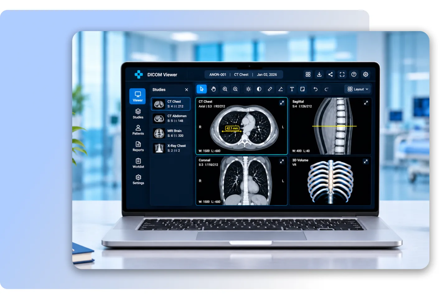
  • Multi-modality supportOpen CT, MRI, X-ray (CR/DX), ultrasound and PET studies in one viewer.
  • High-resolution renderingView images at full resolution with smooth zoom and pan.
  • Window and level presetsSwitch between lung, bone, soft tissue and brain windows in one click, or adjust by mouse.
  • Flexible layoutsCompare series side by side in 1x1, 1x2, 2x2 or custom grids.
  • Cine and stack scrollingScroll through slices or play cine loops for ultrasound and cardiac series.
  • Hanging protocols
  • Series thumbnails and study browserSee every series in a study and drag the one you need into a viewport.
  • DICOM tag viewerInspect the DICOM header of any image when you need the technical details.
Core Viewing in the QSS DICOM/PACS Viewer

Reading, Measurement and Reporting

The tools a radiologist uses on every case, with the report written beside the images.

  • Measurement toolsLength, angle, area, ellipse and region-of-interest measurements with density values on CT.
  • Key imagesFlag the images that matter so referring physicians see them first.
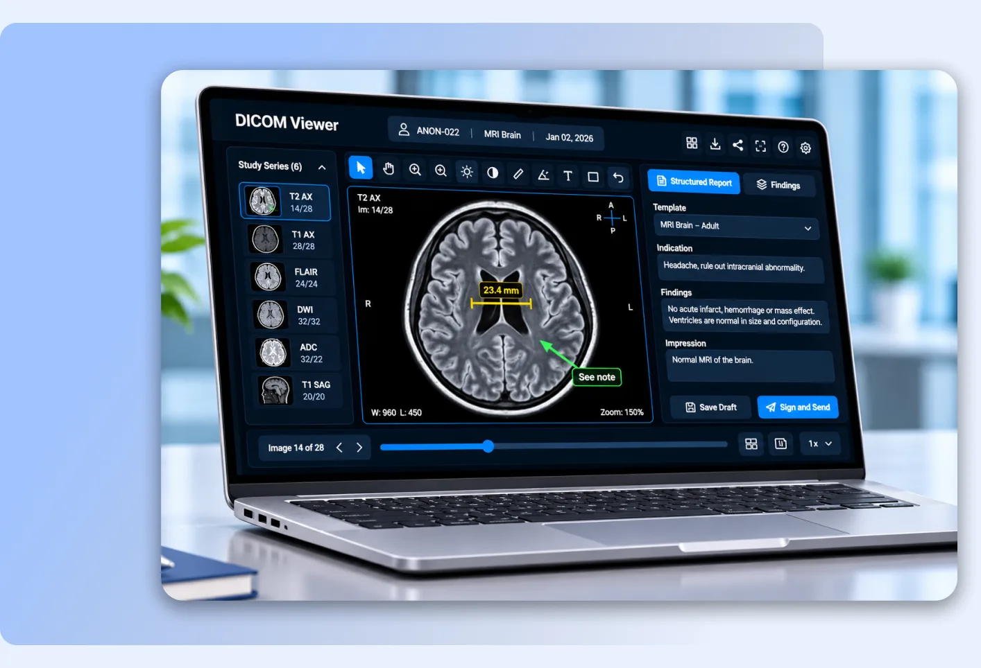
  • Structured reportingUse templates by modality and body part so reports are consistent and complete.
  • WorklistPick up studies from a prioritized worklist filtered by modality, site or status.
  • AnnotationsArrows, text labels and freehand markings saved with the study.
  • Prior study comparisonOpen a patient's previous studies beside the current one with linked scrolling.
  • Report sign-offDraft, review and sign reports, then send them to RIS and EHR.
Reading, Measurement and Reporting in the QSS DICOM/PACS Viewer

Advanced Visualization (MPR and 3D)

A 3D DICOM viewer built into the same workflow, so complex cases do not need a separate workstation.

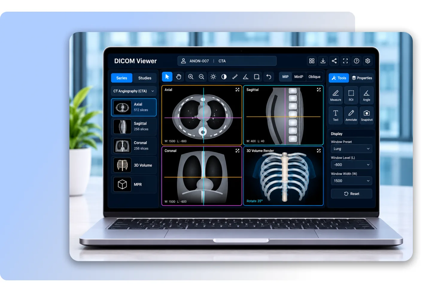
  • Multi-planar reconstruction (MPR)Rebuild axial, sagittal and coronal planes from a single volume.
  • 3D volume renderingTurn CT and MRI volumes into 3D views you can rotate, cut and color.
  • Cross-reference linesSee where the current slice sits on the other planes.
  • PET-CT fusionOverlay functional PET data on anatomical CT for oncology reads.
  • Oblique and curved MPRFollow an angled structure, such as a vessel or the spine, in one plane.
  • MIP and MinIPHighlight vessels or airways with maximum and minimum intensity projections.
  • Synchronized viewportsLink scrolling, zoom and window/level across series and planes.
Advanced Visualization (MPR and 3D) in the QSS DICOM/PACS Viewer

Sharing and Collaboration

Get images to the people who need them, without CDs, couriers or insecure email.

  • Real-time collaborationReview the same study together, with shared annotations, during a consult or tumor board.
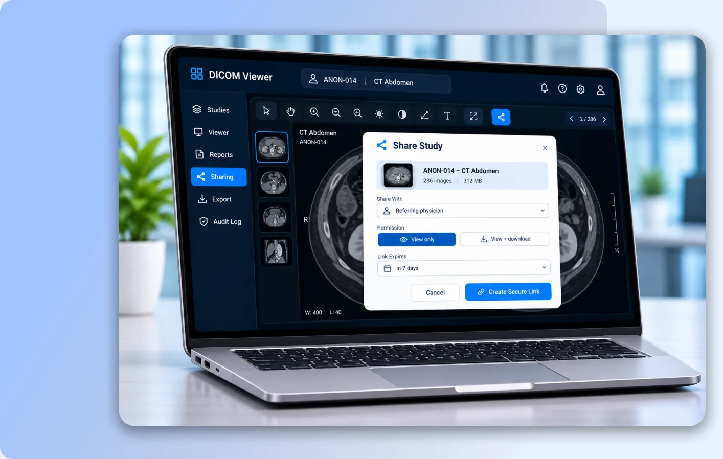
  • Secure study linksShare a study with a referring physician or patient through an expiring, permission-based link.
  • Referring physician accessLet referring doctors view images and reports from a browser, with no install.
  • Patient image accessGive patients a simple way to see and download their own images.
  • CD, DVD and USB exportBurn or copy studies with an embedded viewer for patients and outside providers.
  • DICOM export and sendExport studies as DICOM or send them to another PACS or workstation.
  • Image exportSave selected images as JPEG or PNG for teaching files and presentations.
Sharing and Collaboration in the QSS DICOM/PACS Viewer

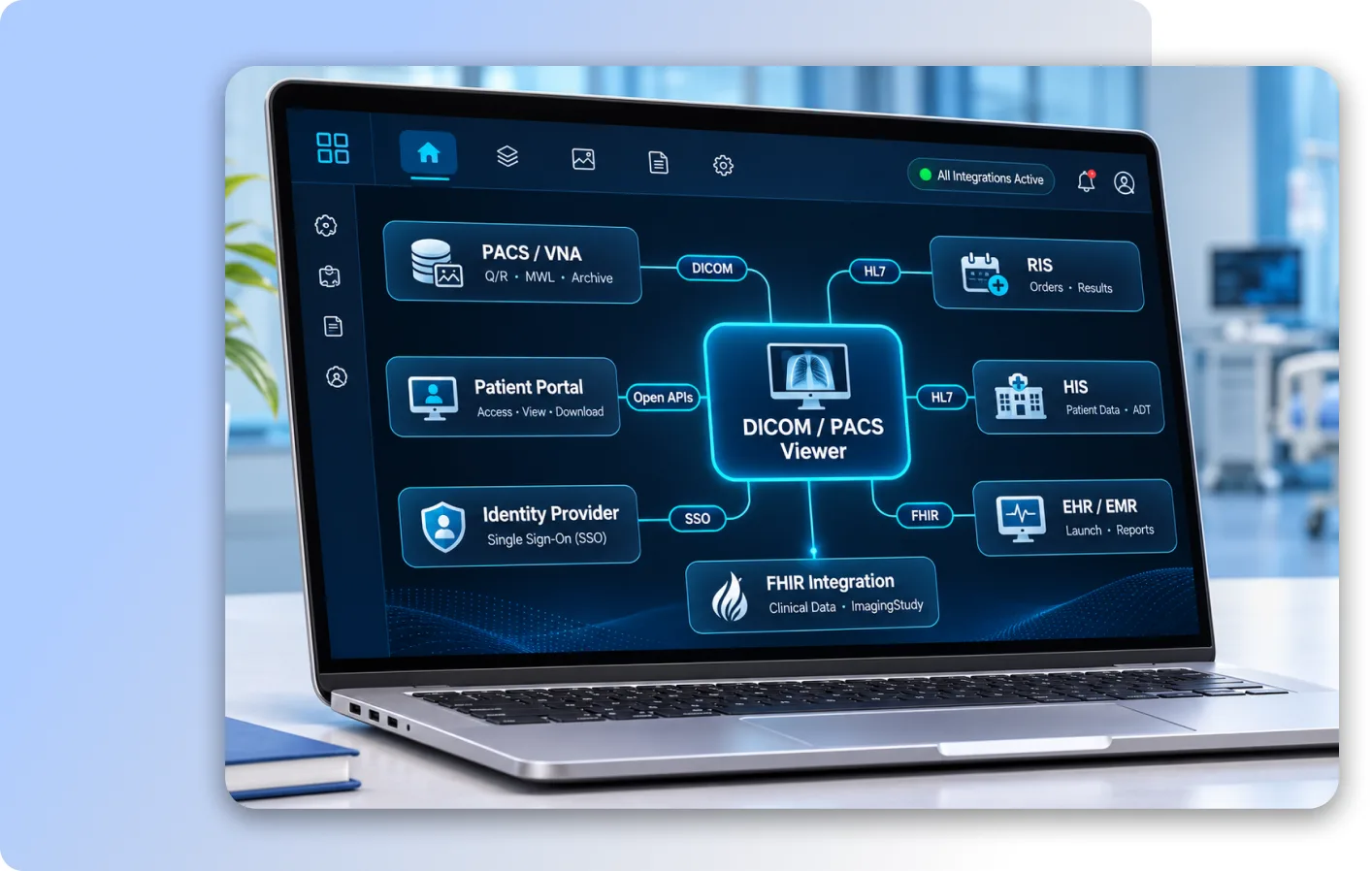
Integration and Interoperability

A PACS viewer that fits into your existing systems instead of asking you to replace them.

  • PACS connectivityQuery and retrieve studies from your PACS and vendor-neutral archive (VNA) over DICOM.
  • DICOMwebUse QIDO-RS, WADO-RS and STOW-RS for fast, web-native image access.
  • Modality worklistWork with DICOM Modality Worklist (MWL) so patient details match the order.
  • RIS and HIS integrationExchange orders, results and patient data over HL7.
  • EHR/EMR integrationLaunch the viewer from the patient chart and return reports to the EHR via HL7 or FHIR.
  • FHIR ImagingStudyExpose study information to FHIR-based apps and portals.
  • Single sign-onSign in with your existing identity provider.
  • Open APIsEmbed the viewer or its tools in your own portal or application.
Integration and Interoperability in the QSS DICOM/PACS Viewer

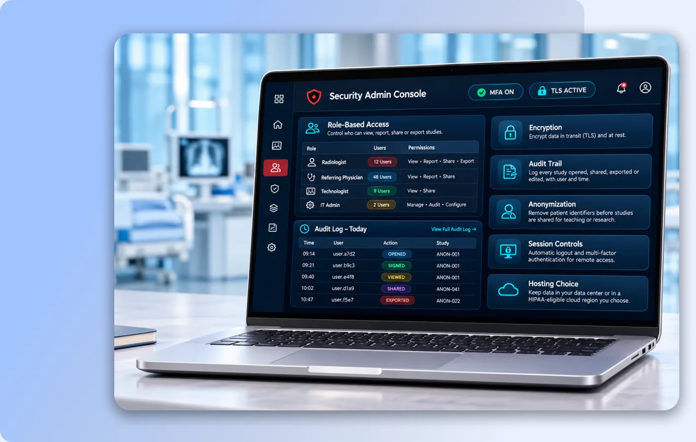
Security and Compliance

Built for HIPAA-regulated environments, by a team certified to ISO 27001.

  • HIPAA-ready designTechnical safeguards that support your HIPAA program for protected health information.
  • Role-based accessControl who can view, report, share or export, by role and by site.
  • EncryptionEncrypt data in transit (TLS) and at rest.
  • Audit trailLog every study opened, shared, exported or edited, with user and time.
  • AnonymizationRemove patient identifiers before studies are shared for teaching or research.
  • Session controlsAutomatic logout and multi-factor authentication for remote access.
  • Hosting choiceKeep data in your data center or in a HIPAA-eligible cloud region you choose.
Security and Compliance in the QSS DICOM/PACS Viewer

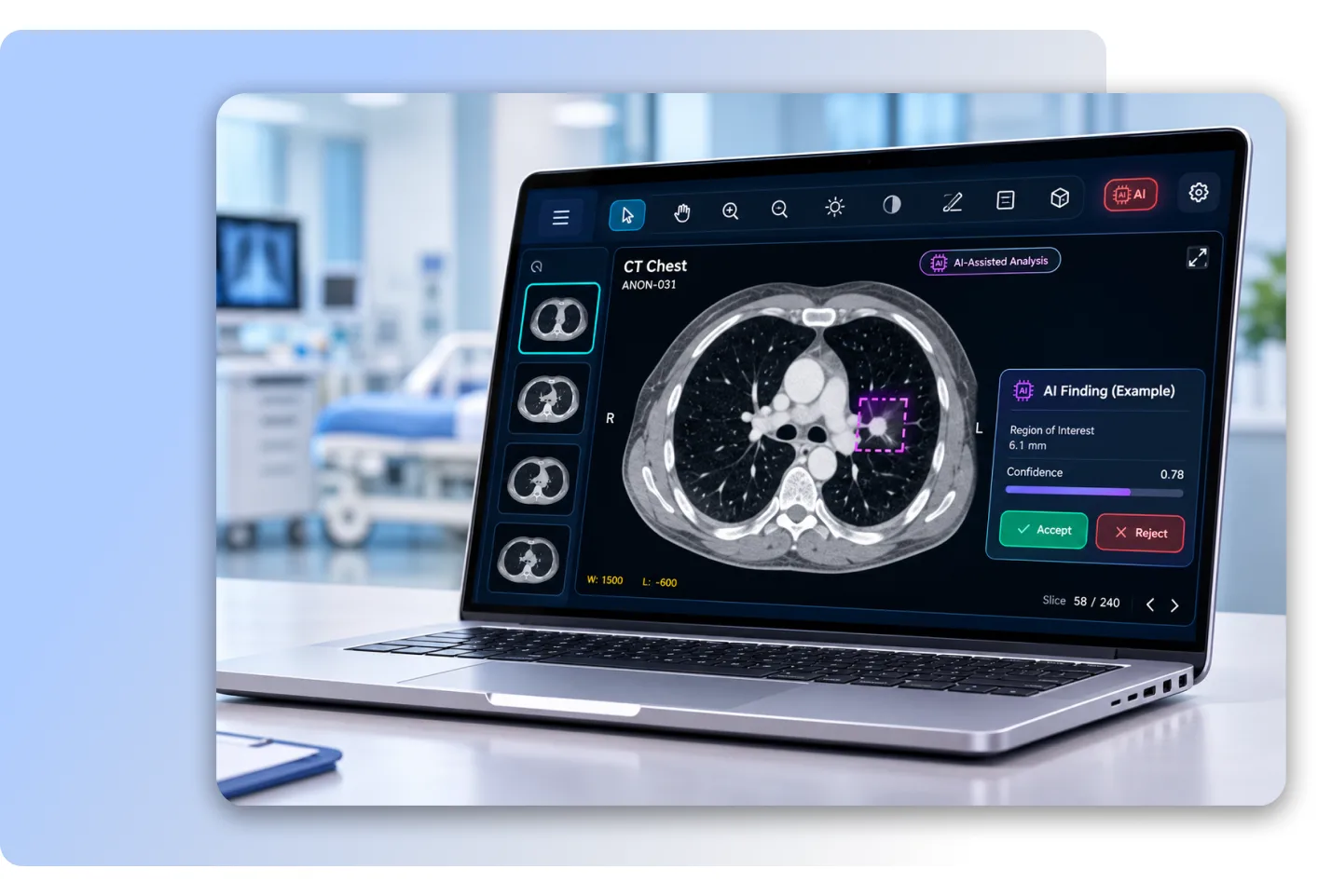
AI-Ready Add-ons

AI tools QSS can integrate into the viewer for your workflow. These are optional add-ons, not built-in diagnostic features.

  • AI result overlaysShow findings from AI models you license as overlays the radiologist can accept or reject.
  • Worklist prioritizationMove studies flagged by an AI triage tool to the top of the worklist.
  • Automated measurementsBring in AI-generated measurements, such as volumes or lesion size, for review.
  • Quantitative image analysisIntegrate quantitative and radiomics tools for research and specialty reads.
  • Model integrationConnect third-party or in-house AI models through standard DICOM and API interfaces.
  • Report drafting supportPre-fill parts of a structured report from AI outputs for the radiologist to edit and sign.
  • Image reconstruction and enhancementConnect AI-based denoising or reconstruction tools to your imaging pipeline.
AI-Ready Add-ons in the QSS DICOM/PACS Viewer

How it works

From scanner to signed report in five steps

Acquire

The modality sends the study to your PACS or archive, matched to the order through the worklist.

Route

The study appears on the radiologist's worklist with priors pulled in automatically.

Read

The radiologist reviews, measures and uses MPR or 3D in the web or desktop viewer.

Report

A structured report is written beside the images, signed and sent to RIS and EHR.

Share

Referring physicians open images and report from the EHR or a secure link; patients get their copy online or on CD/USB.

WHY QSS

Why healthcare teams choose the QSS DICOM/PACS Viewer

You are not just buying a viewer. You get a healthcare software team that builds it, secures it, connects it and supports it for the long term.

01

An experienced medical software development company

For 16+ years, QSS has built healthcare software, from imaging and EHR systems to telehealth and billing platforms, and has delivered 20+ EHR/EMR integrations. We understand clinical workflows, not just code, so the viewer fits how your teams already work.

HL7 interface services →
02

Security by process

Our delivery is CMMI Level 3, ISO 27001 and ISO 9001 certified, and we follow HIPAA-compliant development practices. Access controls, encryption and audit trails are designed in from the first sprint, not added just before go-live.

03

One team for product and integration

The engineers who build the viewer also connect it to your PACS, RIS, HIS and EHR. You work with one accountable team, from setup and integration to custom features and support, instead of coordinating several vendors.

04

Web, desktop and mobile

One product runs in the browser, on reading-room workstations and on phones and tablets, with the same tools and the same study view. Radiologists, physicians and patients each use the device that suits them, with no separate products to manage.

05

Standards first

The viewer is built on DICOM, DICOMweb, HL7 and FHIR, so it works with the PACS, archives and EHRs you already have. Your images stay in standard formats, which keeps you free to change systems later without being locked in.

06

US-headquartered, 24x7 delivery

Your project is led from our headquarters in Bloomington, Minnesota, with delivery centers in the USA, India and Dubai working across time zones. Clients rate us 4.9/5 on Clutch, and 95% of our business comes from repeat clients.

One viewer, the right tools for each role

Everyone who touches a study sees the same images, but each role gets the tools and access it needs, and nothing it does not.

Radiologists

Radiologists

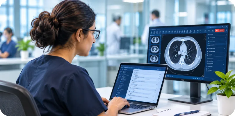
Read faster with hanging protocols, priors side by side, MPR and 3D, and measurement tools in one workspace. Structured report templates turn findings into a clear, consistent report without switching to another system.
Referring physicians

Referring physicians

Open images and the final report straight from the EHR or a secure browser link, with nothing to install. See the key images and measurements the radiologist flagged, so you can act on results sooner.

Specialists and surgeons

Specialists and surgeons

Plan procedures with precise measurements, MPR and 3D volume views in cardiology, orthopedics, neurology and more. Review images before surgery and share annotated views with your team and patients.

Teleradiology groups

Teleradiology groups

Read studies from many hospitals and imaging centers in one web DICOM viewer, from any location. A shared worklist and fast image streaming help remote readers meet turnaround times across sites.
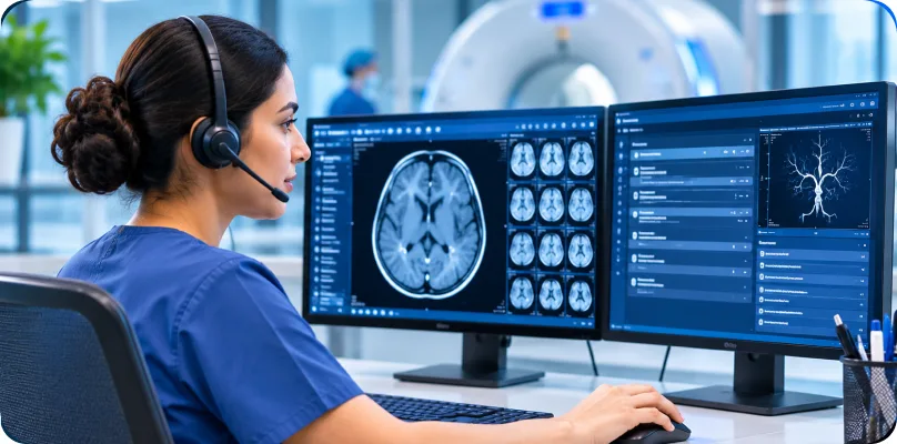
Imaging center and IT leaders

Imaging center and IT leaders

Deploy one viewer across every site, in the cloud, on-premise or hybrid, instead of supporting several tools. Role-based access, audit logs and single sign-on make it easier to secure and manage.
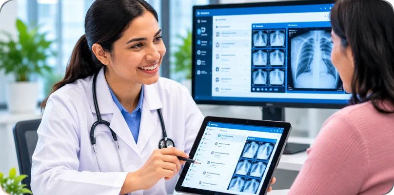
Patients

Patients

View their own images and reports through a simple, secure link or patient portal, on a phone or computer. Share them with another doctor for a second opinion without carrying a CD from clinic to clinic.
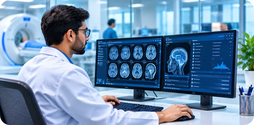
Technologists

Technologists

Check image quality, series and patient details before a study reaches the reading list. Catch missing series or labeling errors early, so radiologists are not slowed down by repeat scans or corrections.

Health tech companies

Health tech companies

Embed a ready DICOM viewer in your own platform, portal or telehealth product instead of building one from scratch. QSS customizes, white-labels and integrates it, so you launch sooner.

Academic and research teams

Academic and research teams

Build teaching files and research datasets from anonymized studies, with annotations and measurements kept. Give students, residents and research partners safe access to cases without exposing patient identity.

Built for the specialties that read images every day

Hospitals and multi-specialty clinics

Give radiology, emergency, surgery and outpatient teams one enterprise viewer across every department and site. Clinicians open the same studies and reports from the EHR, so care teams stay on the same page.

Neurology and neurosurgery

Review brain and spine MRI and CT with MPR, side-by-side priors and precise measurements. Track changes over time and plan surgical approaches with clear 3D views of the anatomy.

Cardiology and vascular

Play cardiac cine loops and review CT angiography with MIP and curved MPR along the vessels. Measure stenosis and vessel size, then share key images with the heart team before procedures.

Orthopedics and sports medicine

Read X-ray, CT and MRI with angle and length tools for alignment, fractures and joint planning. 3D bone views help surgeons explain injuries and plan repairs with patients and their teams.

Gynecology and obstetrics

Review ultrasound studies with cine playback, fetal and pelvic measurements, and annotations. Share selected images and reports securely with patients and referring physicians, without CDs or printed films.

Ophthalmology and optometry

View DICOM images from retinal cameras, OCT and other connected eye-care devices in one place. Compare visits side by side to follow disease progression and share findings with referring doctors.

Diagnostic and imaging labs

Handle high study volumes with worklists, fast image loading and structured reporting templates. Deliver finished reports and images to referring physicians quickly, through the EHR, a portal or a secure link.

Can't find your specialty listed?

Tell us how your team reads images, and we will show you how the viewer fits, or how we can adapt it.

Talk to Our US Team

Industries

Run it where your data needs to be

Choose where the viewer runs and how people reach it. Many sites start on one model and add another later.

Where it runs

CloudOn-premiseHybrid
Your PACSsite
ViewerHIPAA-eligible
cloud
Usersanywhere

Cloud. Hosted in a HIPAA-eligible cloud region, with no servers for your team to manage.

How people use it

Web viewer

Web viewer

A zero-install online DICOM viewer that runs in Chrome, Edge, Safari and Firefox.

Desktop app

Desktop app

A Windows and Mac application for reading rooms and heavy 3D work.

Mobile viewer

Mobile viewer

A mobile DICOM viewer for iOS and Android phones and tablets, for review on the go.

Need it your way? We customize and white-label the viewer

Because QSS builds the viewer, we can change it. Hospitals, imaging networks and health tech companies use it as the starting point for their own imaging product, so they launch faster than building a viewer from scratch.

White-label branding

Your logo, colors and domain, for your clinicians or customers.

Custom tools and workflows

Specialty tools, report templates and hanging protocols built for your team.

Embedding

Put the viewer inside your EHR, portal, telehealth or research platform.

Integrations

Connect to your PACS, VNA, RIS, EHR, billing or AI vendors.

AI integration

Add the AI models you license into the reading workflow.

QSS DICOM/PACS Viewer on a laptop

See it on your studies

Book a live demo of the QSS DICOM/PACS Viewer

In 30 minutes we will show the viewer on anonymized sample studies from your modalities, walk through MPR, 3D and reporting, and discuss how it connects to your PACS, RIS and EHR.

Request your live demo

We reply within one business day.

DICOM Viewer Questions, Answered

Answers about the QSS DICOM/PACS Viewer: what it does, where it runs, how it connects and how it keeps patient data safe.

A DICOM viewer is software that opens medical images stored in the DICOM format, the standard used by CT, MRI, X-ray, ultrasound and PET scanners. It shows the images with their patient and study details and gives readers tools to scroll, adjust contrast, measure and annotate. A PACS viewer does the same while connected to a PACS, the system that stores and distributes those images.

What is the QSS DICOM/PACS Viewer?

What is the difference between a web DICOM viewer and a desktop DICOM viewer?

Which file types and imaging modalities does the viewer support?

Can I use the viewer on a phone or tablet?

People Also Ask
WhatsApp