Problem
Studies take too long to open
 How we solve it
Zero-footprint browser viewers built on OHIF and Cornerstone with progressive loading, streaming and GPU rendering, tuned for large CT and MRI series.

 Reads that start in seconds, on any device.
QSS Technosoft’s medical imaging software development delivers systems that load fast, work in the browser and connect to the record: DICOM viewers, PACS and vendor-neutral archives, teleradiology platforms, RIS/EHR integration and imaging AI.
100+ healthcare projects | 16+ years in healthcare | DICOM · DICOMweb · IHE · HL7 & FHIR
 DICOM • DICOMwebIHE · HL7 & FHIRStandards-based imaging
 100+Healthcare projectsProven healthcare expertise
 CMMILevel 3ISO 27001 · ISO 9001
 16+ yearsIn healthcareUS-headquartered in Bloomington, MN
Trusted Partners
In imaging, speed is the product. As a DICOM PACS development company with a shipped viewer of our own, we build viewers that open studies in seconds and archives that put the image and the report in the record,
for hospitals, radiology groups, teleradiology providers and imaging-AI companies.




CHALLENGES IMAGING TEAMS FACE
Imaging projects rarely fail on the standard. They fail on load time, on the migration that loses priors, on the archive bill and on the AI that never made it into the worklist. Here is where we see imaging organizations get stuck, and how our DICOM and PACS work is built to get them unstuck.
ProblemHow we solve itZero-footprint browser viewers built on OHIF and Cornerstone with progressive loading, streaming and GPU rendering, tuned for large CT and MRI series.
Reads that start in seconds, on any device.ProblemHow we solve itDICOM DIMSE and DICOMweb integration, HL7 v2 ORM/ORU and FHIR ImagingStudy feeds, and IHE SWF and XDS-I profiles, built by the same interface team that does our EHR and HIE work.
Image, order and report in one workflow.ProblemHow we solve itPre-migration audit, header normalization, validated study-level reconciliation and parallel running, so no prior is lost and reading never stops.
A clean archive on day one.ProblemHow we solve itCloud and hybrid archives with lifecycle tiering, compression and a vendor-neutral archive, so the storage decision is yours, not the PACS vendor’s.
Predictable cost per study.ProblemHow we solve itA unified worklist, hanging protocols, priors and AI results inside one viewer, with teleradiology and subspecialty routing.
Audit trail included, not retrofitted.ProblemHow we solve itAI orchestration that runs models on inbound studies and returns results as DICOM SR, overlays or worklist priority, with the audit trail regulators expect.
AI in the workflow, not in a demo. Facing similar challenges?Let’s talk about your reading workflow and show you how to modernize without stopping a single read.Talk to Our US Team →
OUR SERVICES
Six kinds of imaging software, one team. Our medical imaging software development covers the software, the integrations around it and the archive underneath it.
DICOM viewer development
Browser, desktop and mobile viewers for diagnostic and clinical review, built for speed and for the specialty reading them. Start from our own DICOM/PACS Viewer or from OHIF and Cornerstone, and customize from there.
 Zero-footprint web viewersProgressive loading and GPU rendering, nothing to install.
 Advanced visualization2D, MPR, 3D and volume rendering with measurements and annotations.
 Specialty modesRadiology, cardiology, ophthalmology, dental and pathology.
 Embedded viewersInside patient portals, EHRs and referrer access.
 <2 secFirst-image target we design to
 5Specialty reading modes
 3Platforms: web, desktop, mobile
PACS and vendor-neutral archive development
Archives that receive, store, route and serve studies reliably at scale, on-premise, cloud or hybrid, without locking you to one vendor.
DIMSE and DICOMwebC-STORE, C-FIND, C-MOVE, C-GET plus QIDO-RS, WADO-RS and STOW-RS.Vendor-neutral archiveLifecycle tiering, compression and de-duplication.Routing and prefetchingWorklist and hanging-protocol services.Always availableHigh availability, backup and disaster recovery.4DIMSE services3DICOMweb APIs24/7Reading availabilityTeleradiology and enterprise imaging platforms
Multi-site and multi-organization reading with subspecialty routing, secure external access and the reporting tools readers need.
Unified worklistsAcross sites, modalities and referrers.Smart routingSubspecialty and load-balanced routing, peer review and QA.Structured reportingSpeech recognition and templates.Image sharingReferrer and patient access without CDs.1Worklist across every site2Routing modes: subspecialty, load-balanced0CDs needed to share imagesRIS, EHR and modality integration
The connections that put the image and the report in the record: modalities, RIS, EHR, HIS, HIE and billing, on standards rather than one-off workarounds.
Modality connectivityModality worklist (MWL), MPPS and storage commitment.HL7 and FHIRORM, ORU and ADT feeds; FHIR ImagingStudy and DiagnosticReport via Mirth Connect or Iguana.IHE profilesSWF, XDS-I.b, PDI and IOCM.EHR platformsEpic, Oracle Health (Cerner), Meditech and RIS platforms.20+EHR/EMR integrations delivered4IHE profiles supported3HL7 message types: ORM, ORU, ADTDICOM data migration and PACS modernization
Replace or re-platform an archive without losing a prior or stopping reading, with the cost modeled before the first study moves.
Pre-migration auditStudy counts, header quality, orphan and duplicate detection.ValidationHeader normalization, patient-ID reconciliation and study-level checks.Safe cut-overParallel running, phased cut-over and a rollback plan.Cloud migrationAWS HealthImaging, Azure Health Data Services or Google Cloud Healthcare API.4Audit checks before migration3Cloud imaging services supported0Downtime during migrationImaging AI integration and SaMD software
Get models into the reading workflow safely, and build the software around an imaging-AI product so it can pass a hospital IT review and a regulator.
AI orchestrationRun models on inbound studies; return DICOM SR, segmentation and worklist priority.Model integrationThird-party AI marketplaces and in-house models (MONAI, PyTorch, TensorFlow).Dataset toolingAnnotation and de-identification for model training.Regulatory supportDocumentation and verification evidence for FDA and CE pathways.3Output formats: SR, SEG, overlays3AI frameworks supported2Pathways supported: FDA, CEPlenty of firms can describe DICOM. Far fewer have shipped a viewer, migrated an archive without losing a prior, or put an AI result into a radiologist’s worklist with the audit trail a regulator will ask for. QSS has done all three, as a medical software development company that has engineered interoperability and compliance for healthcare clients for 16+ years.

The QSS DICOM/PACS Viewer is in production: web and desktop, multi-modality, structured reporting. Your project starts from a working product, not a blank repository.
Learn more →
We measure time-to-first-image and time-to-full-series on every viewer we ship, and tune streaming, caching and rendering until large series open in seconds.
Learn more →
Dedicated HL7 v2, FHIR, Mirth Connect and Iguana teams; 20+ EHR, EMR and HIS integrations delivered; IHE and DICOMweb by default.
Learn more →
Audited, normalized, reconciled migrations with parallel running and rollback, on-premise or to cloud imaging services.
Learn more →
HIPAA, HITECH, GDPR, PIPEDA and PHIPA; DICOM de-identification and audit; documentation support for FDA and CE pathways where your product is regulated.
Learn more →
Model orchestration, DICOM SR and overlay output, worklist prioritization and the governance that keeps a radiologist in control.
Learn more →The building blocks we draw on most often. Select the ones that matter and we’ll map them to your workflow in the discovery call.
Browser viewer with no install, on any device
Streaming and prefetch so reading starts before the series finishes loading
Multi-planar reconstruction, surface and volume views
Length, angle, ROI, SUV and calcium scoring, saved as DICOM SR/PR
Per-modality, per-specialty layouts with priors
Synchronized side-by-side comparison with earlier studies
CT, MRI, CR/DR, US, PET, NM, MG, OCT, endoscopy and pathology
Unified worklist with subspecialty and load-balanced routing
Templates, speech recognition, key images and DICOM SR
QIDO-RS, WADO-RS and STOW-RS for browser and mobile access
Lifecycle tiering, compression and de-duplication
Run models on inbound studies; return SR, overlays and priority
Echo, angio, ECG and cine playback
Discrepancy tracking and quality workflows
C-STORE, C-FIND, C-MOVE, C-GET, MWL, MPPS and storage commitment
ORM/ORU/ADT feeds; FHIR ImagingStudy and DiagnosticReport
SWF, XDS-I.b, PDI, IOCM and ATNA
Referrer and patient access without CDs, through XDS-I and secure links
Header and pixel anonymization for research and AI
Dataset labeling for model training
AWS HealthImaging, Azure Health Data Services, Google Cloud Healthcare API
Audit, normalize, reconcile and run in parallel
iOS and Android review with secure access
ATNA-style audit, interface and performance monitoring
TLS, MFA, RBAC, encryption at rest and certificate management
HOSPITALS & MULTI-SPECIALTY CLINICS
One imaging system across every department
Streamlined workflows, consolidated patient records and enhanced care coordination.
Learn More → Unified Worklist
 Consolidated Records
 EHR Integration
NEUROLOGY & NEUROSURGERY
High-resolution MRI and CT for brain and spine
High-resolution DICOM tools to visualize MRI and CT scans for faster brain mapping, accurate diagnosis and surgical planning.
Learn More →MRI VisualizationSurgical Planning3D ReconstructionCARDIOLOGY & VASCULAR
Echo, angio and cine at the point of decision
Real-time access to echocardiograms, angiograms and medical images for timely patient care decisions.
Learn More →Echo ReviewCine PlaybackFaster DecisionsORTHOPEDIC & SPORTS MEDICINE
Bone and joint imaging for precise rehabilitation
Manage bone density scans, fractures and joint imaging data for precise rehabilitation plans.
Learn More →Fracture ImagingBone DensityJoint MeasurementsGYNECOLOGY & OBSTETRICS
Secure ultrasound access for women’s health
Secure storage and remote access to ultrasound and fetal monitoring data through cloud-based PACS.
Learn More →Ultrasound StorageFetal MonitoringRemote AccessOPHTHALMOLOGY & OPTOMETRY
OCT imaging built into the eye-care workflow
OCT integration and visualization to support vision diagnostics and ongoing ocular health monitoring.
Learn More →OCT IntegrationVision DiagnosticsOngoing MonitoringDIAGNOSTIC & IMAGING LABS
Faster turnaround from scan to signed report
Faster report turnaround with teleradiology, automated workflows and AI-assisted reading.
Learn More →Faster ReportsTeleradiology ReadingAI AssistanceOrganization Types
 Radiology
 Teleradiology
 AI
 Enterprise
 Dental
 Pathology
 Veterinary
Real results. Real impact. Empowering better care through technology.
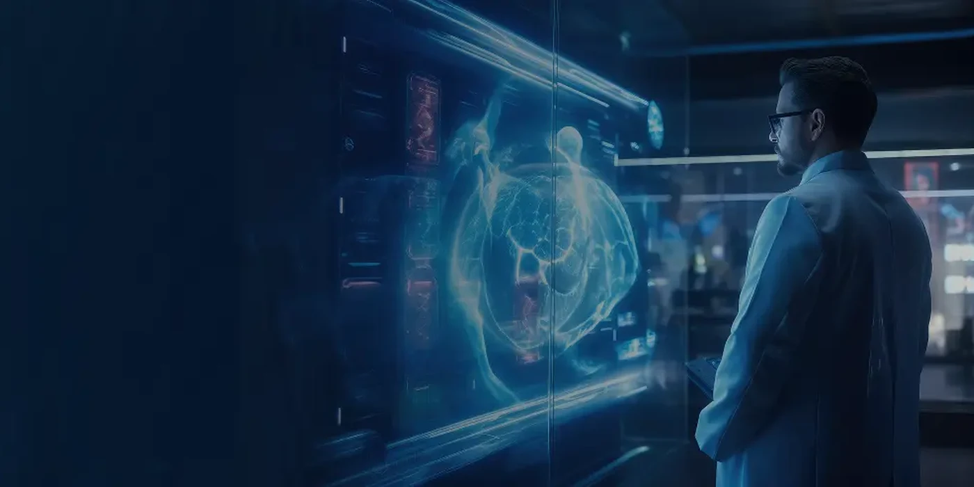
A web and desktop medical imaging viewer built by QSS for radiologists, clinicians and diagnostic specialists. It supports multi-modality DICOM studies, structured reporting, DICOM export and secure patient-data access, and integrates with PACS, HIS, RIS and EHR/EMR systems. Most of our viewer projects start from it.
 Tech Used
DICOM · DICOMweb · HL7 · FHIR · Web and desktop
 Results

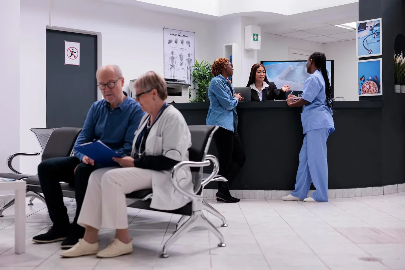
A healthcare connectivity solution bridging hospitals, caregivers, pharmacists and patients in senior living facilities, managing records, prescriptions and care coordination in real time.
Tech Used
.NET Core · Azure · HL7 · SQL Server · Xamarin
Results
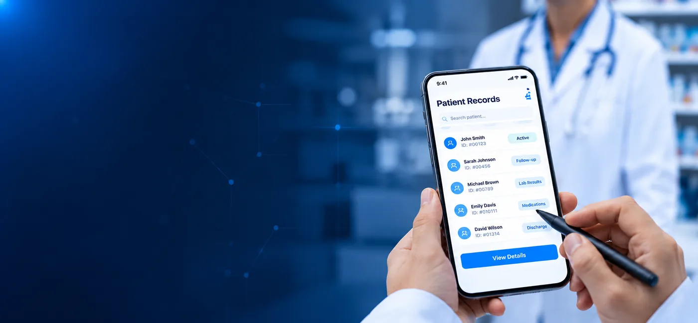
We integrated Iguana to power a cloud-based document storage solution for senior living communities, enabling digital uploads, e-signatures and secure document sharing without physical paperwork.
Tech Used
Iguana · HL7 · REST APIs · AWS · MongoDB · Angular
Results
Most imaging-AI projects stall between the model and the worklist. We build the software that closes that gap and keeps every result reviewable, logged and attributable to a model version.

Imaging Data
New studies, priors,
orders

AI Engine
Detect, segment,
prioritize

Radiologist Review
Human oversight
+ audit trail

Action
Faster, safer
reads
 Worklist prioritizationFlag critical findings and move them to the top of the list
 Segmentation & quantificationOrgan, lesion and vessel measurements pre-filled for review
 Reconstruction & image qualityClean reads from lower-dose acquisitions
 Model operations & governanceVersioning, drift monitoring and a full audit trail
Imaging data is large, identifiable and, when AI is involved, regulated as a device. As a medical software development company, we design every system to the standards and regulations that apply to it, and can show you how, control by control.
AES-256 at rest for archives and TLS 1.2+ for every DIMSE, DICOMweb and API connection, including modality traffic where the device supports it.
MFA, role- and purpose-based access, break-glass with justification, and referrer and patient access scoped to the study.
ATNA-style audit of every study access, export, share and AI result, exportable for OCR audits and internal review.
Header and pixel-level anonymization with configurable profiles for research, AI training and external sharing.
HIPAA-eligible environments on AWS, Azure or Google Cloud with BAAs in place; high availability, backups and tested disaster recovery; on-premise where you require it.
DICOM conformance statements, IHE integration statements and HL7/FHIR interface specifications for every system we ship.
IEC 62304 practices and the documentation and verification evidence your FDA or CE pathway needs. Clearances belong to the product owner.
Third-party vulnerability assessments and penetration tests tracked to closure; load testing on realistic study volumes.

Best when you have a defined viewer, archive or migration scope and a go-live date. Fixed price, milestone-based delivery.

Best when imaging is an ongoing platform. Named DICOM and integration engineers, with front-end and QA included, who work as an extension of your team.

Best when you want to own the capability long term. We build and run the team and platform, then transfer both to you on an agreed timeline.
We choose the stack for your readers and your volumes, not the other way round.
 Imaging standardsDICOM (DIMSE, DICOMweb) · IHE SWF, XDS-I.b, PDI, IOCM, ATNA · HL7 v2 · FHIR R4
 Viewer toolkitsOHIF · Cornerstone3D · VTK.js · ITK · DCMTK · pydicom · fo-dicom · GDCM · OpenGL/WebGL
 Archive & storageOrthanc · dcm4chee · Custom PACS/VNA services · PostgreSQL · S3, Azure Blob, GCS
 Cloud imagingAWS HealthImaging · Azure Health Data Services · Google Cloud Healthcare API
 Interface enginesMirth Connect · Iguana · Rhapsody · Cloverleaf
 AI & computer visionMONAI · PyTorch · TensorFlow · OpenCV · ONNX Runtime · NVIDIA Clara
 Languages & appsC++ · C#/.NET · Java · Python · Node.js · TypeScript · React · Angular · Swift · Kotlin · Flutter
 Security & DevOpsTLS · OAuth 2.0 · SMART on FHIR · MFA · RBAC · Docker · Kubernetes · Terraform
Every imaging system is scoped to its modalities and volumes, but buyers deserve a straight answer. These are typical ranges for medical imaging software development, based on our delivery history and current market rates.

A browser DICOM viewer or one module, such as reporting, sharing or migration tooling
6–12 weeks
$22,000–$40,000
Typical investment

Archive or teleradiology platform with worklists, reporting and integrations
3–6 months
$60,000–$140,000
Typical investment

Multi-site VNA, AI orchestration or a regulated imaging-AI product
6–12+ months, phased
$150,000–$280,000+
Typical investment
Talk to an imaging solutions architect and leave the call with an integration map, a migration risk assessment and a cost range.
 ExpertGuidance
 TailoredRecommendations
 Clear Cost& Timeline
Answers about our medical imaging software development, DICOM and PACS, viewers, migration, imaging AI, compliance, timelines and costs.
DICOM (Digital Imaging and Communications in Medicine) is the global standard for handling, storing, transmitting and viewing medical imaging data. PACS (Picture Archiving and Communication System) is the system healthcare organizations use to store and access images from modalities such as MRI, CT, X-ray and ultrasound. Together they enable image exchange and radiology workflows; DICOMweb extends DICOM to browsers and mobile devices.
Secure image archiving, DICOM and DICOMweb compatibility, zero-footprint web viewers, role-based access control, HL7/FHIR and IHE integration, worklists and routing, structured reporting, mobile access and audit trails for compliance. Increasingly, an AI orchestration layer as well.
We follow HIPAA, GDPR and other healthcare compliance standards. Our PACS solutions use end-to-end encryption, access control, audit logging and secure image-sharing protocols to protect patient data and system integrity; the compliance section above describes each control.
We build tailored viewers for radiologists, cardiologists, orthopedic surgeons, ophthalmologists and others, with 3D reconstruction, annotation and measurement tools, specialty hanging protocols and synchronized comparison with priors.
Yes. We integrate with EHR/EMR, RIS and HIS platforms using HL7 v2, FHIR and DICOMweb, and IHE profiles such as SWF and XDS-I, so imaging orders, images and reports are available from one interface.
System and interface monitoring, performance tuning, user training, feature upgrades, standards and regulatory updates, and 24/7 technical assistance to keep reading uninterrupted.
It improves diagnostic turnaround, puts images and reports in the record, removes manual image handling and CDs, and supports real-time collaboration between radiologists, physicians and specialists. Centralized digital storage, instant access, automated routing and cloud options reduce both delay and cost.
Yes. Cloud and hybrid PACS on AWS HealthImaging, Azure Health Data Services or Google Cloud Healthcare API offer scalability, remote access, automated backups and lower infrastructure cost, and suit multi-location groups and teleradiology. We model the storage and egress cost before anything is provisioned.
Yes. We integrate AI/ML models into viewers and archives for worklist prioritization, segmentation, quantification and reconstruction, returning results as DICOM SR, SEG and overlays, with model versioning and audit so a radiologist stays in control.
All DICOM-compliant modalities, including MRI, CT, PET, CR, DR, ultrasound, nuclear medicine, mammography, OCT, endoscopy and whole-slide pathology.
We build vendor-neutral archives and use DICOM, HL7, FHIR and IHE profiles (XDS-I, SWF, PDI) to ensure cross-platform compatibility and image sharing across departments, facilities and external partners.
Because we are a medical software development company that has shipped a DICOM/PACS Viewer of our own, migrated archives without losing priors and put AI into reading workflows, all on an interoperability practice with 20+ EHR, EMR and HIS integrations behind it. Common PACS implementation challenges (legacy integration, migration, multi-site scale, compliance, adoption, specialty workflows, vendor lock-in) are the things we plan for at discovery rather than discover at go-live.
Medical imaging software development covers viewers, archives, integration, migration and AI around DICOM data. It differs from ordinary software in three ways: the data is large and identifiable, so performance engineering and privacy controls are the core work, not polish; the users are readers whose time is measured in seconds per study; and the software may be regulated as a medical device, so documentation and verification evidence are part of the deliverable. A DICOM PACS development company that has shipped its own viewer plans around all three from day one.
Yes. We audit the current archive, normalize headers, reconcile patient IDs and validate at study level, then run the new archive in parallel and cut over by site or modality with a rollback plan. Reading never stops, and no prior is lost.
Yes. As a medical software development company, we build the viewer, orchestration, integration and cloud software around an imaging-AI product to IEC 62304 practices, and we produce the documentation and verification evidence the pathway needs. Clearances belong to the product owner; we help you get there.
We set time-to-first-image and time-to-full-series targets at discovery and measure them every sprint on your own de-identified studies. For typical CT and MRI series on a browser viewer with progressive loading, first image in under two seconds is the target we design to; your network and archive placement decide the rest, which is why we model both before building.
Yes. Most projects start from our own DICOM/PACS Viewer, but we also integrate leading third-party viewers such as RadiAnt when a workflow requires it, so new software fits the tools your readers already use.
Yes. Our viewer can save and export studies to CD, DVD or USB for offline sharing, and the DICOM export keeps files standards-compliant during transfer. For routine sharing, we recommend secure links and XDS-I, which remove the need for physical media.